
-Custom GUI to control 6 skin options, including full color / gray scale / x-ray, on / off toggles for each system, and transparency sliders for each system. Built-in controls also enable the model to spin 360 degrees without interfering with the lighting. -Every item is selectable and correctly titled / categorized for simple organizing. -Is it possible to isolate any muscle, even deep tissue muscle, and observe its proper origin and insertion into the underlying skeleton? -Real World Scale -Native Maya File -Textures that are realistic -Extremely detailed displacement map -Squeaky-clean edge -topology based on loops (no n-gons, minimal triangles) -Lighting setup and ready to render -79cm in height, 72cm in width, and 31cm in depth TEXTURES ================== (4096x4096) Arm Color.tif (4096x4096) Arm Displacement.tif (4096x4096) ConnectiveTissue Color.tif (4096x4096) ConnectiveTissue NormalMap.tif (2048x1024) hdrImageBasedLighting.hdr (4096x4096) Leg Color.tif (4096x4096) Leg Displacement.tif (4096x4096) Arm Muscles.tif (4096x4096) Face Color of Muscles.tif (8192x8192) FemaleTorso Muscles Color.tif (4096x4096) Color Muscles Leg.tif (4096x4096) Pelvis Color.tif (4096x4096) Pelvic Displacement.tif (8192x8192) Ribs Color.tif (8192x8192) Torso Skeleton Color.tif (8192x8192) Skin Female Color.tif (4096x4096) Skin Female Displacement.tif
The profile is the next stage in learning how to sketch a body. Begin by sketching the head again, this time in the form of an egg but with the end pointing diagonally down, and then drop a vertical line from the crown to the ground. In an upright position, the pelvic bone (a smaller version of the head's egg), the shoulder, and the knee may all be found nearly on this vertical line. All of the joints are on the same level as previously, but the others are not on the same plane as these.
Now that we've covered the musculature, let's look at how it relates to the pelvis. The pelvis is basically a fundamental component that binds the torso's internal organs together. Try to envision the pelvis in relation to the muscles and organs above it with gravity applied. Here are two examples of fictitious exercises.
You have full right, power, legal ability, and authority to enter into and fulfill this Agreement, have acquired any necessary third-party permission, and have had a chance to seek independent legal guidance prior to any Purchase. You will only use Digital Assets in accordance with the provisions of this Agreement. If you use Digital Assets in an unlawful manner, you agree to pay RenderHub any reasonable cost or penalty imposed by this Agreement or relevant law. You will assess the need for and, if necessary, get any necessary third-party permission, approval, or release to use Other Intellectual Property displayed in a Digital Asset prior to Purchase, and you will not use a Digital Asset to violate any partyâs Other Intellectual Property rights. Before revealing any rights problem to a third party, you shall promptly inform RenderHub of any legal claim or challenge to your use of a Digital Asset. VIII. LIABILITY LIMITATION
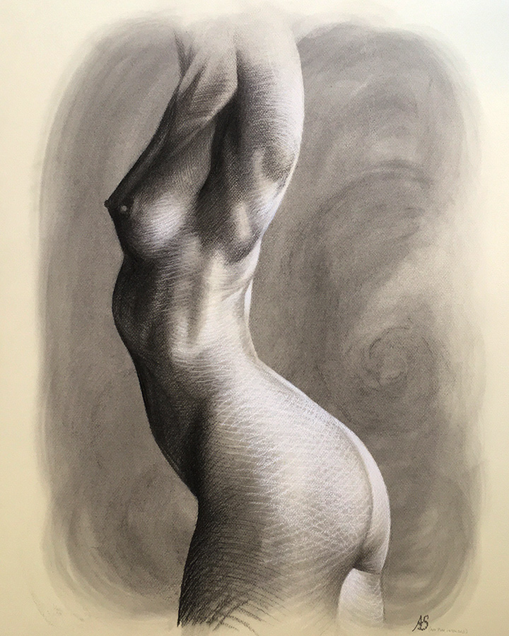
Female Torso Anatomy Diagram
During menstruation, the endometrium grows and develops in preparation for the embryo as the ovum matures and passes through the fallopian tube. If the ovum is not fertilized in time or fails to implant into the endometrium, the uterine arteries constrict, cutting off blood supply to the endometrial. Menstruation is caused by a lack of blood flow in the endometrium, which results in cell death and tissue loss. This shedding occurs around day 28 of a typical menstrual cycle and continues throughout the first few days of the following reproductive cycle.
==ADVANCED FILES== To improve compatibility, an OBJ file has been included. Please keep in mind that the OBJ file comprises the whole model (hierarchy free) with all Color Maps allocated as well as a.mtl file. The use of advanced xRay shaders, control rigs, and displacement maps is not permitted. To improve compatibility, an FBX file has been included. Please keep in mind that the FBX file has the whole model (with hyerarchy and layers intact) as well as all Color Maps allocated. Some applications may not support advanced xRay shaders, control rigs, or displacement maps.
With the noteworthy exception of the brain, most essential organs in humans are contained inside the torso. The rib cage protects the heart and lungs in the upper chest, and the abdomen contains the majority of the digestive organs: the stomach, which breaks down partially digested food via gastric acid; the liver, which produces bile required for digestion; the large and small intestines, which extract nutrients from food; the anus, from which fecal wastes are egested; the rectum, which stores feces; and the gallbladder, which stores and concentrates Finally, both the male and female reproductive organs are housed in the pelvic area. Muscle groups of importance [Correction]
As previously stated, we all have a layer of fatty tissue under our skin. It may be very thin in our most ripped athletes and completely absent in starving bodies, but it is there in even healthy bodies — in fact, it is what helps us conceive of a body as healthy, as opposed to "skin and bones." Furthermore, fat reserves are kept in different regions of the body, and they are not the same in men and girls! Females store fat in their underarms, thighs, and buttocks, while men store fat in their bellies.
Female Chest Anatomy Diagram
The goblet cell and bronchial seromucous glands produce mucous containing immunoglobulin A, cytokines, and other cytolytic substances, as well as extensive mucosal production of nitric oxide by the nose and paranasal sinuses; all of these have potent antimicrobial activity, protecting underlining tissues and trapping organisms and particles. Bacteria in the mucus are killed by lysozyme. Lymphocytes, which occupy the lamina propria in huge numbers, also provide extra protection against microorganisms. Large blood veins in the lamina propria assist to warm the air. [8] Furthermore, the inferior concha has a very extensive venous plexus known as the erectile tissue. Every 30 to 60 minutes, erectile tissue on one side engorges and inhibits air flow through that fossa, forcing air to be diverted via the other nose and fossa, giving the engorged side time to recover from drying. Every hour or so, the flow of air alternates between the right and left nostrils. Inflammation increases mucous production (for example, in asthma and bronchitis), and illnesses may change its composition. [8]
This is a frequent source of vaginal irritation, and according to the Centers for Disease Control and Prevention, at least 75% of adult women have had one at some point in their lives. Candida, a fungus that overgrows in the vagina, causes yeast infections. Yeast infections are often caused by an imbalance in the vaginal pH, which is typically acidic. Other causes include pregnancy, diabetes, compromised immune systems, tight-fitting garments, and douching. Itching, burning, irritation, and a white cottage-cheese-like discharge from the vagina are all symptoms of yeast infections. Women have experienced painful intercourse and urination as well. A yeast infection may be diagnosed by taking a sample of vaginal secretions and examining them under a microscope for indications of yeast. Treatment options range from lotions that may be placed in or around the vaginal region to oral medications that inhibit fungal development. [4] [edit] Genital mutilation
Lactation is the process of producing and releasing milk to nourish a newborn. Milk production occurs before to birth and is regulated by the hormone prolactin. Because prolactin is created in response to a newborn sucking on the nipple, milk is produced as long as vigorous breastfeeding is practiced. Prolactin and milk production cease as soon as a baby is weaned. The milk-letdown reflex, which is mediated by the hormone oxytocin, is the release of milk by the nipples. Because oxytocin is also created in reaction to newborn suckling, milk is only released when the infant is actively eating.
The main (oblique) fissures extend obliquely forward and downward from the fifth thoracic vertebral body to the diaphragm, dividing the lungs into upper and lower lobes (Fig. 1-9). The right major fissure is more oblique, finishes more anteriorly (on the lateral chest radiograph), merges with the right hemidiaphragm, and connects the minor fissure. The minor (horizontal) fissure divides the right middle lobe from the right upper lobe and spreads forward and laterally from the right hilus (Fig. 1-10). It is uncommon to be able to trace both fissures on chest radiographs in their entirety. Fissures are often "incomplete," merely partly separating lobes. The location of the main cracks is frequently seen on CT images as a belt of avascularity. Depending on slice thickness, the fissure may be imperceptible or apparent as a poorly defined or well-defined band of density. On CT scans, the site of the minor fissure may be extrapolated from the vast oval deficit of arteries on one or more sections at the bronchus intermedius level.
Female Abdominal Anatomy Diagram
depicts the interior anatomy of a female abdomen in historical art. Huge selection, incredible variety, 100+ million high quality, inexpensive rf and rm photos. Anatomytools.com offers extremely realistic male and female anatomical reference models, artist busts, instructional dvds, armatures and workshops utilized by fx artists, 3d artists, medical professionals and sculptors. Female and male anatomy female: Female abdominal müscles are anatomy female genital. This hd wallpaper female abdominal anatomy photos has seen by 1012 people.
https://www.verywellhealth.com/female-body-diagram-5209032
Nov 29, 2021 · Vagina: The vagina is a muscular tube that links the cervix and the uterus, going to the outside of the body. Parts of the vagina are rich in collagen and elastin, which give it the potential to expand during sexual stimulation and birthing. Cervix: The cervix is the bottom section of the uterus that divides the lower uterus and the vagina and may have a role in lubrication.


